


Hip joint coxarthrosis (HJ) is a degenerative-dystrophic disease that affects cartilage and bone tissue. In medical articles, it can be called differently: deforming coxarthrosis, DOA of the hip joint, osteoarthritis. All these terms mean the same pathology - osteoarthritis, but "coxarthrosis" is a narrower concept that characterizes the loss of the hip joint.
Cartilage is the first to suffer from osteoarthritis, then the bones and surrounding structures - ligaments and muscles - are involved in the pathological process. If there are changes in the bones, the prefix "osteo" is added to the word "osteoarthritis". In advanced cases the joint is deformed and already talk about deforming osteoarthritis (osteoarthritis).
general characteristics
Deforming osteoarthritis of the hip joint is the second most common after knee gonarthrosis. Due to the deep location of the thigh joint, the bone deformation can go unnoticed for a long time and only X-ray images taken at later stages will show changes.
The development of this disease is influenced by various factors, including an inactive lifestyle, trauma and metabolic disorders. It is because of the specifics of modern life, in which there is often no room for physical education, that osteoarthritis affects an increasing number of people. Moreover, the peak incidence falls in the middle age group - from 40 to 60 years.
Reference:coxarthrosis often affects women more than men.
Development mechanism
The thigh joint is formed by two bones: the femur and the iliac (pelvis). The femoral head is inserted into the pelvic acetabulum, which remains immobile during movement - walking, running. At the same time, the articular surface of the femur can move in several directions, providing flexion, extension, abduction, adduction, and rotation of the thigh.
During physical activity, the femur moves freely in the acetabulum due to the cartilaginous tissue that covers the articular surfaces. Hyaline cartilage is distinguished for its strength, durability and elasticity; acts as a shock absorber and participates in the distribution of load during human movements.
Inside the joint is the synovial fluid - the synovium - which is essential for lubricating and nourishing cartilage. The entire joint is enclosed in a dense and thin capsule surrounded by strong muscles in the thighs and buttocks. These muscles, also acting as shock absorbers, serve to prevent damage to the thigh joint.
The development of coxarthrosis begins with changes in the joint fluid, which becomes more viscous and thicker. Due to the lack of moisture, the cartilage does not get enough food and begins to dry out, it loses its softness and cracks appear on it.
The bones can no longer move as freely as before, and they rub against each other, causing micro-damage to the cartilage. The pressure between the bones increases, the cartilage layer becomes thinner. Under the influence of increasing pressure, the bones gradually deform, local metabolic processes are disrupted. In the later stages a pronounced atrophy of the leg muscles is observed.
Causes
Deformative arthrosis of the thigh joint can be primary and secondary. It is not always possible to determine the cause of primary osteoarthritis. Secondary arthrosis appears against the background of existing diseases, namely:
- congenital hip dislocation or hip dysplasia;
- Perthes disease (aseptic necrosis of the femoral head);
- hip joint coxarthritis, which has an infectious, rheumatic or other origin;
- pelvic bone injuries - dislocations, fractures.
Hip dysplasia is a congenital malformation that sometimes does not appear clinically for a long time and in the future (aged 25-55 years) may lead to the development of dysplastic coxarthrosis.
Coxo arthrosis can be left, right and symmetrical. In primary osteoarthritis, concomitant diseases of the musculoskeletal system are often observed - in particular, osteochondrosis and gonarthrosis.
There are also risk factors that contribute to the onset of the disease:
- excess weight and excessive loads overloading the joints;
- violation of blood circulation and metabolism;
- hormonal shifts;
- curvature of the spine, flat feet;
- old age;
- hypodynamics;
- inheritance.
It should be noted that coxarthrosis itself is not inherited. However, some features of metabolism or connective tissue structure may create preconditions for the development of osteoarthritis in a child in the future.
Symptoms of coxarthrosis
The main symptom of osteoarthritis of the thigh is pain in the groin and groin area, which has different intensity. Stiffness and stiffness during movement are also noticed, a decrease in muscle volume, shortening of the affected limb and a change in gait due to lameness.
Coxarthrosis most often progresses slowly, causing initial discomfort and mild pain after exertion. However, over time, the pain increases and appears at rest.
A typical manifestation of pathology is difficulty in grabbing the thighs, when a person can not sit "on foot" in a chair. The presence and severity of signs of coxarthrosis depends on its degree, but pain syndrome is always present.
There are three degrees or types of ankle osteoarthritis, which vary in the severity of the injury and the accompanying symptoms:
- 1 degree. The thigh does not hurt all the time, but periodically, mainly after walking or standing for a long time on foot. The pain syndrome is localized to the ankle area, but can sometimes spread to the legs up to the knee. Muscles with grade 1 coxarthrosis do not decrease in size, gait does not change, motor ability is fully preserved;
- 2nd degree. Feelings of pain intensify, arise not only after running or walking, but also at rest. The pain is most often concentrated in the thigh area, but can spread to the knee. In moments of heavy load, it is painful to step on the injured limb, so the patient begins to spare the leg and lame. The range of motion in the joints is reduced, it is especially difficult to move the leg sideways or rotate the thigh;
- 3 degrees. The pain becomes permanent and does not go away even at night. The gait is visibly impaired, the independent movement is visibly complicated, and the patient leans on a cane. The range of motion is severely limited, the muscles of the buttocks and the whole leg, including the lower leg, atrophy.
- Due to muscle weakness, the pelvis bends forward, the affected leg is shortened. To compensate for the difference in limb length, the patient tilts the body to the affected side when walking. This leads to a shift in the center of gravity and increased stress on the affected joint.
Osteoarthritis or osteoarthritis?
Arthritis is an inflammation of the joint, which can be an independent disease or develop against the background of systemic pathologies (for example, rheumatism). In addition to the inflammatory response, symptoms of osteoarthritis (especially in the advanced stages) include limited mobility and changes in the shape of the joint.
At the heart of degenerative-dystrophic changes of osteoarthritis is damage to cartilage tissue, which often leads to the appearance of inflammation. This is why osteoarthritis is sometimes called osteoarthritis. And since osteoarthritis is almost always associated with joint deformity, the term "osteoarthritis" is applicable to it.
Reference:according to the International Classification of Diseases (ICD-10), osteoarthritis and osteoarthritis are varieties of the same pathology.
Diagnosis of coxarthrosis
The diagnosis of "hip joint coxarthrosis" is made on the basis of examination, patient complaints, and examination results. The most informative method is radiography: in the picture you can see both the degree of damage to the joint and the cause of the disease.
For example, in hip dysplasia, the acetabulum is flatter and sloping, and the cervico-diaphyseal angle (inclination of the femoral neck in the vertical plane) is larger than normal. Deformity of the femoral part located near the ankle is characteristic of Perthes disease.
Grade 3 coxarthrosis is characterized by narrowing of the ankle space, enlargement of the femoral head, and numerous bone growths (osteophytes).
If the patient has had a fracture or dislocation, signs of trauma will also be visible on X-rays. If a detailed assessment of the condition of the bones and soft tissues is required, magnetic resonance imaging or computed tomography may be prescribed.
Differential diagnosis is performed with the following diseases:
- gonarthrosis;
- osteochondrosis and radicular syndrome arising in its background;
- trochanteritis (inflammation of the trochanter bone of the thigh);
- ankylosing spondylitis;
- reactive arthritis.
The decrease in muscle volume that accompanies stages 2 and 3 of coxarthrosis can cause pain in the knee area. Moreover, the knee often hurts even more than the hip joint itself. An x-ray is usually enough to confirm the diagnosis and rule out gonarthrosis.
With diseases of the spine - osteochondrosis and tight nerve roots - the pain is very similar to coxarthrosis. However, it happens suddenly, after an unsuccessful movement, a sharp turn of the body or lifting a weight. Pain sensations start in the gluteal region and spread to the back of the leg.
Radicular syndrome is characterized by severe pain when a straight limb is raised from a supine position. However, there is no difficulty in abducting the leg sideways, as with coxarthrosis. It is worth noting that osteochondrosis and osteoarthritis of the hip joint are often diagnosed simultaneously, so a comprehensive examination is needed.
Trochanteritis, or trochanteric bursitis, develops rapidly, unlike osteoarthritis, which can progress slowly over years and even decades. The pain syndrome develops within a week or two, while it is quite intense. The cause of trochanteritis is trauma or excessive exercise. Movement is not restricted and the leg is not shortened.
Ankylosing spondylitis and reactive arthritis can also be associated with symptoms that mimic coxarthrosis. The distinguishing sign of such diseases is the appearance of pain mainly at night. The groin can hurt quite badly, but when you walk and move, the pain subsides. In the morning, patients are concerned about the hardening, which disappears after a few hours.
Treatment of hip arthrosis
Coxarthrosis can be treated conservatively or surgically. The choice of treatment method depends on the stage and nature of the course of the pathological process. If diagnosed with grade 1 or 2 disease, it is treated with medication and physiotherapy. After relieving the acute symptoms, therapeutic exercises and massage are added to them. If necessary, a special diet is recommended.
The sooner coxarthrosis is detected and treated, the more favorable the prognosis. With the help of medicines and therapeutic measures, you can significantly slow down the pathological process and improve the quality of life.
Non-steroidal anti-inflammatory drugs (NSAIDs) are used to relieve pain and inflammation. It should be noted that anesthesia is performed in the shortest possible course, as NSAID-class drugs can adversely affect the digestive tract and slow down the regeneration processes in cartilage tissue.
It is possible to accelerate cartilage restoration with the help of chondroprotectors. However, these funds are effective only in the early stages of the disease, when its hyaline cartilage is not completely destroyed. Chondroprotectors are prescribed in the form of tablets or intra-articular injections.
Vasodilators are used to improve blood supply to the joints. For muscle spasms, muscle relaxants are advised.
In the case of persistent pain syndrome, which is difficult to eliminate with pills, injections are made into the hip joint. Corticosteroids relieve inflammation and pain well.
Medication therapy can also be supplemented with topical agents - ointments and gels. They do not have a pronounced effect, but help to cope with muscle spasms and reduce pain.
Physiotherapy helps improve blood circulation and cartilage nutrition. For coxarthrosis, procedures like shock wave therapy (SWT), magnetotherapy, infrared laser, ultrasound and hydrogen sulfide baths have proven to be excellent.
operation
Treatment of stage 3 osteoarthritis can only be surgical, as the joint is almost completely destroyed. To restore the function of the hip joint, partial or total arthroplasty is performed.
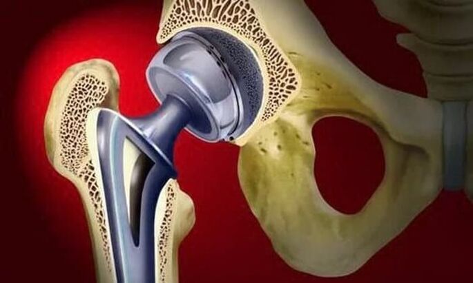
Surgical treatment is used in advanced cases of osteoarthritis, when conservative therapy is powerless.
In partial prosthesis, only the femoral head is replaced with an artificial prosthesis. Total prosthesis means replacement of the femoral head and acetabulum. The operation is performed under general anesthesia and in the vast majority of cases (about 95%) the function of the thigh joint is fully restored.
During the rehabilitation period, antibiotics are prescribed to the patient to prevent infectious complications. Sutures are removed on day 10-12 and exercise therapy begins. The attending physician helps the patient learn to walk and distribute the load correctly on the operated limb. Exercise is an important step to increase muscle strength, endurance and elasticity.
Work capacity returns on average 2-3 months after surgery, but for the elderly this period can be up to six months. Upon completion of rehabilitation, patients can fully move, work, and even play sports. The service life of the prosthesis is at least 15 years. To replace a worn prosthesis, a second operation is performed.
effects
Without timely and adequate treatment, coxarthrosis can not only significantly worsen the quality of life, but also lead to disability and disability. Already in the second stage of osteoarthritis, the patient is given the third group of disability.
When you cut the affected limb by 7 cm or more, when a person moves only with the help of improvised means, a second group is assigned. The first group of disability is accepted by patients with third degree coxarthrosis, accompanied by complete loss of motor ability.
Indications for medical and social examination (MSK) are:
- long course of osteoarthritis, more than three years, with regular irritations. The frequency of irritations is at least three times every 12 months;
- has undergone an endoprosthetic operation;
- severe disorders of musculoskeletal function of the limbs.
Prophylaxis
The main measures to prevent coxarthrosis are diet (if you are overweight) and regular but moderate physical activity. It is very important to avoid damage to the pelvic region and hypothermia.
In the presence of risk factors for the development of osteoarthritis, as well as all patients with a diagnosed disease, swimming is beneficial. Sports such as running, jumping, football and tennis are not recommended.