


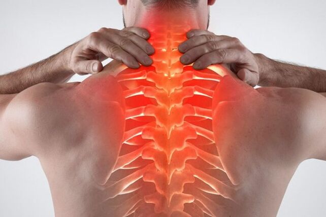
Chest osteochondrosis is a common degenerative disease. There are specific symptoms of thoracic osteochondrosis, indicating the onset of pathology. In the initial stages, the concern does not bother the patient much, so he does not rush to seek help from a specialist. Over time, the symptoms intensify, which forces the patient to go to the doctor, where a neglected pathology is found. You need to find out which early signs of osteochondrosis are defined and which treatment methods are most effective.
What is thoracic osteochondrosis and how does it arise?
Osteochondrosis of the thoracic region is characterized by the appearance of destructive-dystrophic processes in the middle part of the ridge. The destruction is located between the 8th and 19th beads. To find out which vertebra is affected, it is necessary to perform accurate diagnostic studies. Osteochondrosis of the thoracic region is often associated with frightening complications, including prolapse or hernia. Without complications, the disease is rare, as the destruction of cartilage tissue inevitably leads to the destruction of the entire vertebral frame.
When a patient develops a circulatory disorder or age-related ear joint, the fibrous ring located in the intervertebral disc cavity begins to collapse, losing its normal structure. Since the destruction is slow, then in the initial stages microcracks appear, through which the nucleus penetrates.
As the internal component comes out, the anal fibrosis begins to weaken, leading to gradual stretching and rupture. When the pulposus nucleus emerges, an intervertebral hernia occurs, which is the most common complication of osteochondrosis. Pathology involves cartilage tissue damage, which causes significant concern. Severe back pain is also associated with neurological syndromes that develop from the bite or irritation of nerve roots.
Symptoms of breast osteochondrosis
In the initial stage, the patient does not feel discomfort, therefore, at this stage, the disease can be detected only by chance. The disease has many symptoms that can be disguised as other pathologies.
Symptoms of chest osteochondrosis can be felt from the following manifestations:
- Breathing is difficult. Problems arise, manifested by shortness of breath and the feeling of shortness of breath. This indicates damage to the thoracic vertebrae and spinal cord.
- The main symptom is pain in the chest area. There is also a pressing feeling in the heart, rather reminiscent of an ischemic attack.
- Discomfort occurs when the back bends. As the disease progresses, the pain in this position increases.
- Against the background of deteriorating blood circulation, there is a feeling of coldness in the lower or upper extremities.
- Chest pain on the background of intervertebral hernias appearing. The discomfort is often felt most strongly on the left or right side of the affected area.
- Throat discomfort and swallowing problems. If there is irritation of the nerve endings in the upper thoracic region, a cough appears.
- Women may experience chest pain unrelated to cyclical changes or hormonal imbalance.
- The sensation of tingling or burning sensation appears in the area of the feet and legs.
- Hair and nails become brittle, dull.
- Shingles occurs less frequently.
- Back and chest pain occurs at the same time.
- Rarely, there are concerns in the stomach, liver or pancreas.
- Onset of severe rib pain, indicating intercostal neuralgia.
- There are signs of chondrosis and chest compression - a similar pathology.
- There are problems in the work of the gastrointestinal tract. Feels nauseous, heaviness in the stomach.
- In men, some lusts may fall. Problems arise in the genitourinary sphere.
- When you stand or sit for a long time, severe discomfort appears.
- There is a strong headache accompanied by dizziness. Migraines with aura may occur.
- The patient often develops intercostal neuralgia.
- The pain may radiate to the neck or lower back.
If you find in total thoracic osteochondrosis and its signs or some of them, it is necessary to urgently consult a therapist, neurologist, orthopedist. Also, such symptoms should be reported in the absence of problems with the gastrointestinal tract, cardiovascular system and lungs.
There are also acute and subacute symptoms. If, with a worsening of osteochondrosis in the chest region, the patient experiences severe pain that deprives the patient of the ability to work, and he can only observe bed rest, then the subacute course is slow and does not significantly limit the patient's motor activity.
A clear sign of a slow injury - no acute pain. Symptoms in the subacute phase disappear. No discomfort with basic body movements, including sucking, sneezing or turning. A person does not suffer from pain in a dream, so the process of falling asleep is facilitated.
So that the subacute course of the disease does not worsen and pass into remission, important rules must be observed:
- Lifting weights is prohibited.
- You can not bow sharply.
- It is forbidden to be in a sitting or standing position for a long time. A person often unconsciously in this condition assumes a detrimental posture, so there is an excessive load on the back, which brings on another deterioration.
- Avoid hypothermia. Beenshtë proved that non-observance of a comfortable temperature regime for the body turns into a worsening of the inflammatory process. Humidity is also harmful to the joints.
The duration of the subacute course is individual. If you follow the medical recommendations, the patient will completely get rid of the discomfort within 2-3 weeks. If conservative treatment and rest do not help, and the patient begins to suffer from nausea, dizziness and weakness, an urgent need to consult a specialist. Such symptoms indicate a re-deterioration.
Degrees of development of osteochondrosis in the chest region
There are 4 clinical stages of the disease, during the onset of which the patient develops signs of pathology:
- In the initial stage, there are no clinical symptoms. The first stage occurs against the background of the appearance of destructive processes in cartilage and bone tissue. In the first stage, there is also no rupture or stretching of the fibrous ring, so there is no hernia either. They can detect the initial elongation and signs of cartilage degeneration.
- The second stage presents with slight pain or discomfort. An attentive patient seeks a physician, therefore, osteochondrosis of the chest region is detected immediately. People who do not want to visit a specialist can still endure the second stage, using the available remedies, but self-medication will not be enough for a long time. At this stage, the most common neurological symptoms may appear, including headache, burning in the intercapsular area, neck pain, and hypertension. Also at this stage, there is an increase in degenerative destruction in the spine: the fibrous ring comes out, which leads to the appearance of an intervertebral hernia of the thoracic region.
- The third stage is already difficult for the patient. Persistent neurological syndromes develop, including persistent radiating pain in the shoulder blades, arms, bone marrow, and lower back. The patient may exhibit somatic and autonomic disorders, indicating a disturbance in the functioning of the nervous system. The patient is often tortured by nausea, incessant headaches, dizziness, back pain. Masked cardiac, gastroenterological, or pulmonary signs of disease may also appear. At this stage, there is an active demineralization of bone and cartilage tissue. There is a tendency for injury.
- The last stage is the fourth. Against the background of osteochondrosis and hernias, irreversible consequences arise - the mobility of intervertebral discs is completely lost, and cartilage tissue at the site of a long course of inflammation is replaced by osteophytes. An operation is required to remove them.
In order not to run the body in a state similar to stage 3 or 4, it is better to visit a doctor at the smallest sign. The sooner the disease is detected and therapy is started, the sooner the patient will return to normal and learn to live with osteochondrosis. The destructive pathological process can not be completely stopped, but can be slowed down by leading a healthy lifestyle, using medication and performing daily gymnastics. The later the patient returns to the doctor, the more difficult it is to stop the severe pain syndrome associated with cartilage tissue degeneration.
Risk factors and causes of the disease
There is no exact reason that causes devastating changes in the spine. An important role in the occurrence of pathology is attributed to an inherited factor. Beenshte proved that individuals who suffer from physical inactivity are more likely to have crest problems than those who exercise regularly. Excessive physical activity can also provoke cartilage destruction at a young age.
Thinning and destruction of intervertebral discs is closely related to spinal overload. If the muscles are not strong enough, and the back is subjected to regular overload, destruction of cartilage tissue occurs.
What causes osteochondrosis:
- Mbipesha. When you are overweight, there is a strong weight pressure on your back. As a result, premature destruction of bone tissue occurs.
- The presence of an abnormality in the structure of bone and cartilage. Such problems also arise during the period of intrauterine development.
- Congenital asymmetry of intra-articular gaps in the intervertebral nodes of the type of tropism anomaly, contributing to the emergence of a degenerative-dystrophic process in the spine.
- Presence of muscle spasm, spondylosis, chronic persistent trigger points, and vascular disorders in the chest region. These pathologies also contribute to the occurrence of osteochondrosis of the chest region.
- Prolonged exposure to vibrations in the spine in a sitting position. An example of a job is a minibus or bus driver.
- Frequent physical exertion accompanied by heavy lifting. Examples are workloads or professional sports activities.
- Smoking and alcohol abuse. People with healthy lifestyles are more likely to have mineral deficiencies in their bodies and poor circulation, leading to back problems.
- Sedentary lifestyle. With insufficient physical activity, an accelerated rinsing of calcium occurs, which is associated with poor metabolic processes. As a result, the bones become brittle. Also, muscle tissue atrophies, due to which the load on the spine increases greatly. The result is pain, frequent discomfort with minimal physical exertion.
Due to the intervertebral discs, sufficient crest mobility is ensured. Intervertebral discs play a shock-absorbing role. With the development of osteochondrosis, an accelerated process of demineralization occurs, vital moisture from the joints is lost. This leads to discomfort, reduced mobility in the spine.
Risk factors for breast osteochondrosis include:
- Advanced age. In older people, natural degeneration occurs, therefore, after 40 years, the disease is detected more often.
- Female. In girls, there are periods that contribute to the active rinsing of calcium from the bones - pregnancy and menopause. Without adequate pharmacological support, spinal diseases are prone to occur.
- Presence of hormonal disorders, endocrinological diseases. If the patient has diabetes mellitus or uncompensated hypothyroidism, degeneration of the intervertebral disc can occur at an early age.
- Prolonged immobilization. If the patient is ill and has to lie down for a long time, atrophic processes occur in the muscles, which causes back pain.
- Previous back injuries. When ligaments and tendons are stretched, the risk of osteochondrosis in the chest region increases.
- Presence of scoliosis. Poor posture in the future provokes serious spinal problems, including osteochondrosis and hernia.
Diagnosis of thoracic osteochondrosis
If the patient suspects back problems, it is necessary to consult a therapist. The doctor conducts a general examination of the patient, asks about complaints, measures blood pressure. If a neurological problem is suspected, the patient is referred to a specialist - a traumatologist, neurologist or orthopedist.
In the meeting with a specialized specialist, they also ask about complaints, perform an initial diagnosis of the patient. Based on a visual examination, a range of diagnostic measures are described, including:
- Radiography. With the help of an X-ray, you can assess the condition of the skeletal system in general terms. If the patient has a hernia or osteochondrosis, signs of pathology may be noticed - the distance between the intervertebral discs will be reduced and darkening is sometimes observed at the alleged site of the hernia. If the image results do not suit the specialist, you should continue to look for the cause of the pain and discomfort.
- CT or MRI. The most accurate diagnostic methods that allow you to accurately examine the state of the focus of inflammation in the picture. A more detailed image can be seen on MRI, but if there are contraindications (presence of a pacemaker or prostheses in the joints), computed tomography is prescribed. CT is an enhanced X-ray that allows you to see bone, tendon and ligament in detail. The image makes the image in the form of a three-dimensional image, so the details of the damage are clearly visible.
- Biochemical and general blood test. These tests are needed to assess the patient's health. If an increase in leukocytes, ESR, is detected, then this indicates an active inflammatory process in the body. With active destruction of bone tissue, reduced calcium levels and a deficiency of cholecalciferol (vitamin D3) are found in the blood.
- Spine scintigraphy. The research method reveals active destruction of bone tissue. Weak bone tissue is very sensitive to fragility. The method will detect the tendency and signs of degeneration.
To diagnose the disease, you should consult an experienced specialist. For the final diagnosis, a complete clinical picture is needed, taking into account some laboratory research methods.
Thoracic spine osteochondrosis requires differentiation along with the following pathologies:
- Dichormonal spondylopathy.
- Urinary tract pathologies, including urolithiasis, cystitis or pyelonephritis.
- Diseases of the cardiovascular system, excluding sinus arrhythmia, tachycardia and angina pectoris.
- Gastrointestinal tract diseases, including chronic pancreatitis, gastric and duodenal ulcers, irritable bowel syndrome.
- Previous injuries, fractures.
- Tumors in the chest, including a malignant flow.
- Rheumatoid arthritis (determined by a blood test for C-reactive protein, rheumatism test and ESR).
- Spinal osteomyelitis.
- Acute inflammatory process.
- Ankylosing spondylitis.
- Spondylolisthesis.
Treatment of osteochondrosis of the thoracic spine
An integrated approach to therapy is needed to slow the progression of the disease. In the initial stages, only conservative therapy is indicated, which consists of the use of medications and physiotherapy treatment methods. In advanced cases, when the patient has large hernias and a pronounced degree of bone degeneration, an operation is prescribed. Do not treat yourself at home. Folk remedies do not eliminate osteochondrosis of the thoracic spine.
In which cases is the operation performed?
Initiated osteochondrosis of the thoracic region adversely affects the patient's quality of life. If the patient has persistent disturbances that interfere with normal life, given the lack of effect of medication treatment, then a surgical solution to the problem may be offered.
Absolute indications for surgery include:
- Lack of sensitivity in the bladder and intestines.
- If the tenderness in the foot disappears and the patient loses the ability to move independently.
- Paralysis due to strong hernia growth.
In other cases, the patient makes the decision to remove the hernia formation independently. If the disease really brings severe anxiety and the patient's condition does not improve against the background of conservative treatment, doctors recommend surgery.
Drug treatment of thoracic spine osteochondrosis
During the period of deterioration, the attending physician prescribes various medications necessary for use in order to facilitate the inflammatory process. The acute period is characterized by severe pain that can only be relieved with medication. If enough medicine is taken, the patient improves. Only an experienced specialist can prescribe medication; self-medication is unacceptable.
Osteochondrosis of the thoracic spine is treated with the following medications:
- Non-steroidal anti-inflammatory drugs, pain relievers, or analgesics. These medications are designed to quickly relieve back pain accompanied by an active inflammatory process. The effect of taking pills or injections is felt the next day. Taking any medicine from the NSAID group is associated with side effects with prolonged use, therefore, experts recommend limiting the use of the drug to the minimum period, not more than 1-2 weeks. Painkillers are more harmful to the gastric mucosa, causing gastropathy and inflammation. Patients at risk are given certain medications designed to protect the gastrointestinal mucosa. Examples are proton pump inhibitors, histamine H2 receptor blockers, antacids. People with ulcers and gastritis are better off avoiding the use of NSAIDs or taking modern analogues with a selective effect.
- Muscle relaxants. These drugs are very effective in treating muscle spasticity. Relieve pain associated with muscle tension. They act on the trigger points located in the crushed muscle tissue. The more a person is overloaded, the higher their number. Muscle relaxants relieve muscle tightness well, and therefore exhibit an analgesic effect. You should take medication in a course, the average duration of therapy is at least 2-4 weeks.
- Group B vitamins Assign B1, B6, B12 in the form of injections with a combined composition. In large doses, these substances have an analgesic effect and have a positive effect on the nervous system. Neurotrophic drugs are effective in treating pain associated with suppressed nerve roots. With the help of nutrition, it is impossible to meet the norm of these substances needed to achieve a therapeutic effect, therefore they are prescribed in the form of medicines. The average length of a course of injections is 2-3 weeks. Then, if necessary, they switch to tablets.
- Anti-inflammatory ointments, gels. If pain is tolerable, and systemic forms of NSAIDs are contraindicated, external medications are prescribed. The advantage of external remedies is that they do not cause side effects. In rare cases, skin allergies may occur, but the ointment will not cause gastrointestinal or laboratory blood deterioration. Another advantage of outdoor products is the possibility of long-term use. You can rub in gels for up to 4 weeks, after which they take a break. The scheme and duration of therapy is determined by the attending physician.
- Honroprotectors. These are complex substances used to nourish the articular cartilage tissue. It is necessary to use medication for a long course, at least six months, after which they take a break of 2-3 months and the course of therapy is repeated. Within 2-3 months release injection forms are used, as they are better absorbed. Then they switch to supportive treatment, including the use of tablets. Importers It is important to understand that the medication does not stop the destruction of cartilage tissue. They create only extra food, which slows down the degenerative processes that occur in the bones and joints.
- Complex preparations of calcium and vitamin D3. Beinshte proved that the inhabitants of the northern latitudes do not get enough vitamin D3 because solar activity is low all year round in this region. To get rid of hypovitaminosis, it is necessary to take cholecalciferol supplements in winter and autumn in courses while solar activity is minimal. Without this vitamin, the assimilation of calcium and other minerals is impossible. Due to a long calcium deficiency, thinning of bone tissue occurs over time, so a person suffers from osteochondrosis and other complications. Calcium and D3 are better absorbed in combination, therefore complex preparations are described. The dose and course of administration should be prescribed by the attending physician.
As an adjunct to treatment, homeopathy, antispasmodics and complex multivitamins may be prescribed.
Conservative therapy for breast osteochondrosis
During the recovery period, the patient should pay sufficient attention to rehabilitation. The more carefully the patient maintains health, the less disease attacks will occur.
The most effective conservative treatments include:
- Exercise therapy. With the help of exercises, the patient learns to keep his back straight, strengthens muscular corsets. Physiotherapy can be done at any age, several times a week. The complex is chosen individually, taking into account the anatomical features of the patient. Start the execution gradually, initially spending no more than 5 minutes a day. As physical qualities improve, the patient learns to do more strenuous exercise over a longer period of time.
- Supporting corset. Anatomical devices serve to support weakened muscles, if there are contraindications to strengthening them. The patient chooses a bandage depending on the height and type of appointment. The attending physician must select the appropriate model. The duration and pattern of the dress is determined individually. You can not wear a corset all the time, otherwise your back muscles will become even weaker.
- Massage. In medical practice, massages are one of the most popular and at the same time effective methods of conservative treatment, in the presence of osteochondrosis of the thoracic region in a patient. During the recovery period, the muscles need extra support. Useful is useful when blood flow is temporarily improved and the overstretched muscles are immobile using the proper technique. You should attend specialist sessions several times a year for courses.
- Physiotherapy. Physiotherapy procedures are prevalent in traumatic, orthopedic and neurological practice. With the help of procedures, local blood flow is improved, systemic medications are used externally and the apparatus acts on damaged tissues. As a result, the muscles warm up and the chronic inflammatory process is eliminated in the affected area. Examples of medical procedures - magnetotherapy, shock wave therapy, electrophoresis.
Rarely, manual therapy and acupuncture are prescribed.
Osteochondrosis of the thoracic region is a serious disease if it starts. To prevent the disease from proceeding acutely, it is necessary to treat the pathology comprehensively.